Synthesizing porous fluorescent silica nanoparticles for analyte sensing in the brain
Since Spring 2022
This is my graduation thesis of spring 2023 and is a collaboration of the Adair lab and the Gluckman lab.
Our objective is to study the real-time changes in concentration of analytes such as extracellular potassium and oxygen in the brain.
To do reach our goal we are using a fiber endoscope that we developed last year.
![]()
The endoscope uses LED light to ratiometrically measure through diodes the real time changes in light wavelength at the fiber stub (which is the end that enters the brain).
Currently I am developing the sensor responsible for the change in light signal and to do so I am making a coating of porous silica nanoparticles with ion K+ and O2 sensitive fluorophores encapsulated in them
Porosity
During Summer 2022 I was awarded the Erickson Grant to work on my thesis and I focused on making the particles the most porous possible.
The porosity is a crucial step to determine the quality of the sensor, since a higher porosity means that more ions can enter the particle and interact with the fluorophore, permitting a more sensitive detector and a more precise analysis. The challenge is not only to have the highest possible porosity but also to have microporous (with pores < 2nm) particles so that the ions can penetrate the particles and interact with the fluorophores inside, keeping the larger fluorophores trapped in the smaller pores.
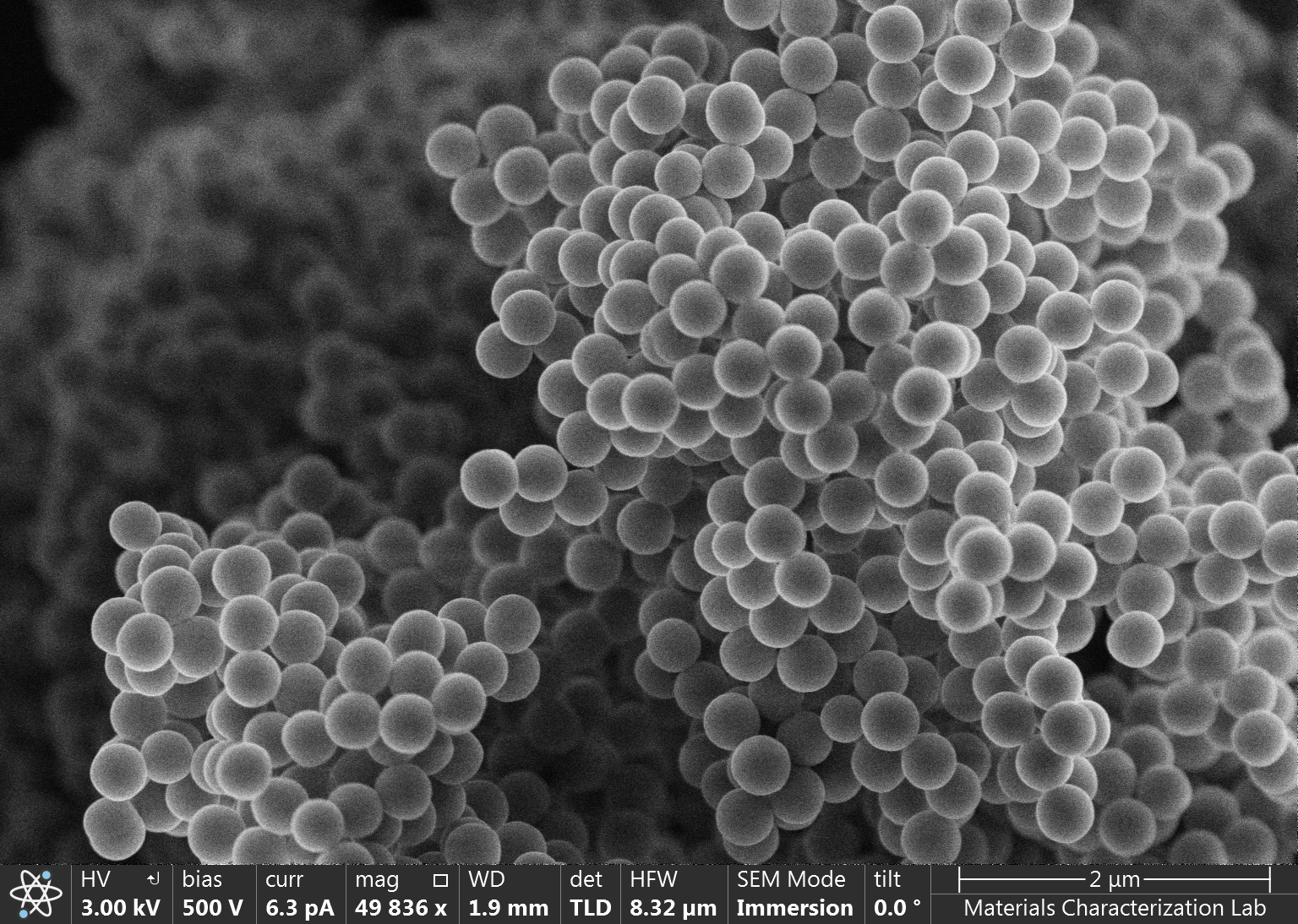
To create porosity in the particles, we decided to use a revised Stober method using TMOS in different molar percentages compared to TEOS. TMOS by having the methyl group rather than the ethyl group, hydrolyzes much faster, making the aggregation process so rapid that continuous porosity is formed in the nanoparticle structure.
![]()
![]()
![]()
![]()
![]()
Our objective is to study the real-time changes in concentration of analytes such as extracellular potassium and oxygen in the brain.
Design
To do reach our goal we are using a fiber endoscope that we developed last year.
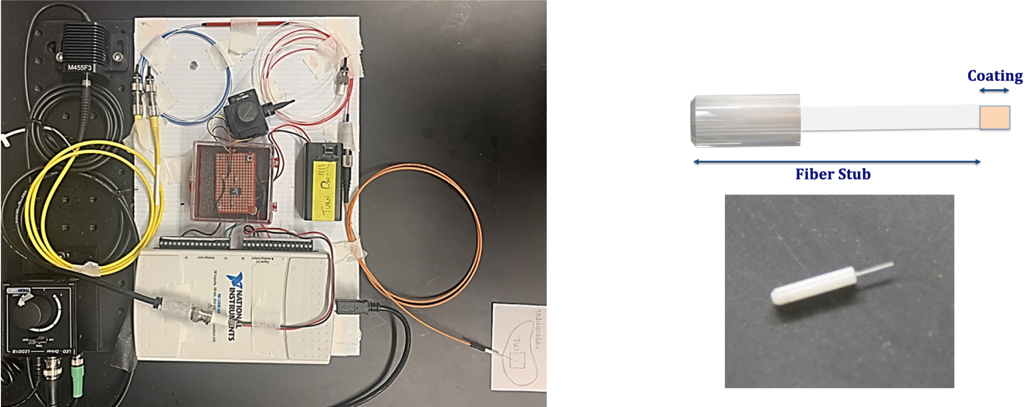
Image from: Gangarosa R. (2022). Utilizing Fiber fluorescence for oxygen sensing in living tissue. The Pennsylvania State University
The endoscope uses LED light to ratiometrically measure through diodes the real time changes in light wavelength at the fiber stub (which is the end that enters the brain).
Currently I am developing the sensor responsible for the change in light signal and to do so I am making a coating of porous silica nanoparticles with ion K+ and O2 sensitive fluorophores encapsulated in them
Porosity
During Summer 2022 I was awarded the Erickson Grant to work on my thesis and I focused on making the particles the most porous possible.
The porosity is a crucial step to determine the quality of the sensor, since a higher porosity means that more ions can enter the particle and interact with the fluorophore, permitting a more sensitive detector and a more precise analysis. The challenge is not only to have the highest possible porosity but also to have microporous (with pores < 2nm) particles so that the ions can penetrate the particles and interact with the fluorophores inside, keeping the larger fluorophores trapped in the smaller pores.
To create porosity in the particles, we decided to use a revised Stober method using TMOS in different molar percentages compared to TEOS. TMOS by having the methyl group rather than the ethyl group, hydrolyzes much faster, making the aggregation process so rapid that continuous porosity is formed in the nanoparticle structure.
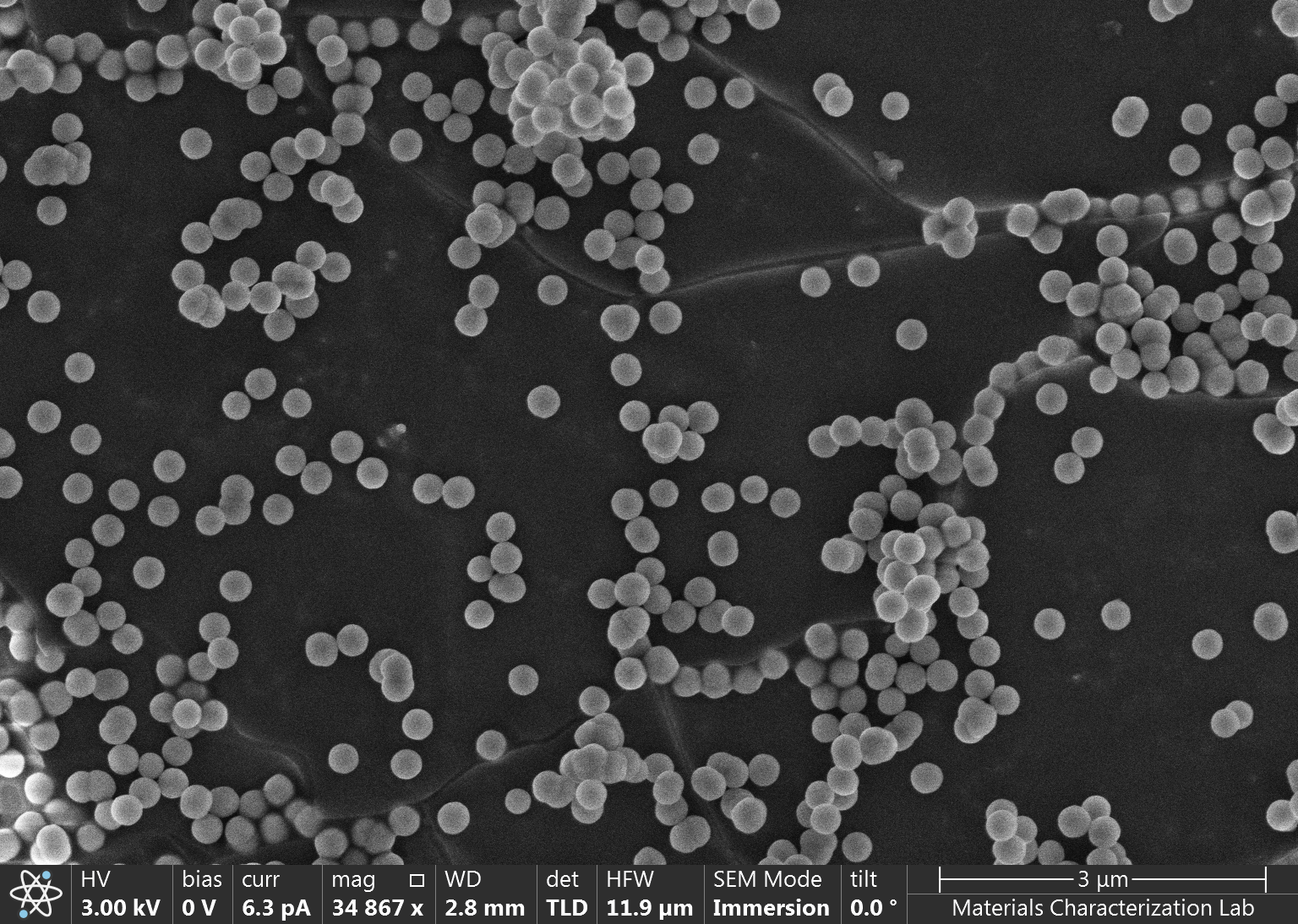
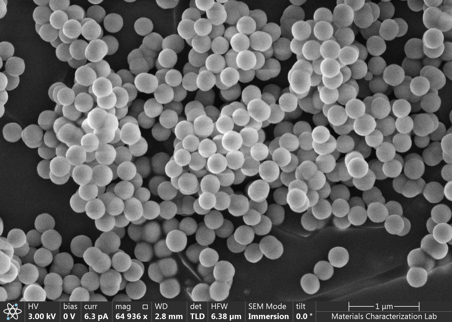


On the left, from the top to the bottom is shown the behavior of the particles’ surface area, particle diameter, pore size, porosity and normalized surface area over different molar percentages of TMOS. On the right is shown the SEM images gathered at different percentages of the fabricated porous silica nanoparticles.
Right now
Right now we are investigating the best way to include the fluorophores in the reaction and we are observing how their properties are changed by being encapsulated in the particles. Once that is defined, we will proceed in connecting the particles to the fiber stub and test the endoscope in different environments and conditions.Advantages
This system compared to others is simpler, cheaper, customizable and has a better spatial and temporal resolution.
The encapsulation of the Nanoparticles has three core advantages:
- restrict distribution of the extracellular space
-
lower the photobleaching rate of the fluorophores
-
allow co-localization of fluorophores with reporter fluorophores for quantitative ratio metric measures and FRET excitation.
